Corneal Topography

Table of Contents
Corneal Topography System
Corneal topography System is a specialized imaging technique that maps the surface of the cornea, the clear, front part of the eye. Similar to a 3D map showing terrain features, it identifies irregularities in the normally smooth corneal curvature. This test helps detect distortions, monitor eye diseases, and assist in surgical planning.
The procedure is quick, painless, and non-contact, generating color-coded maps that aid in diagnosing and managing various eye conditions. Corneal topography is essential for pre-operative planning for procedures like LASIK and other eye surgeries.
What is Corneal Topography?
Corneal topography is a painless diagnostic test that creates color-coded maps of your cornea, the transparent, outer layer of your eye. The cornea has a curved shape that bends (refracts) light to help you see clearly. By analyzing the shape of your cornea, corneal topography helps identify and manage various eye conditions.
The term “topography” typically refers to surface features of land, such as mountains or rivers, and the maps that illustrate them. In a similar way, your cornea can have a smooth surface or contain subtle irregularities. While these features aren’t visible to the naked eye, corneal topography maps them by measuring the cornea’s shape, thickness, and elevation changes.
This test is the gold standard for detecting subtle corneal changes, whether they develop suddenly or gradually. It is also referred to as computerized corneal topography and is essential for diagnosing eye conditions and planning treatments like LASIK surgery.
What Conditions Can Corneal Topography Diagnose and Monitor?
- Scarring: Corneal trauma or infections can lead to scarring, which alters the cornea’s shape. A topography scan measures these distortions and their impact on vision.
- Growths: Corneal topography can track the size and progression of pterygia or other growths.
- Astigmatism and Keratoconus: This scan helps detect astigmatism and early stages of keratoconus, while also monitoring their development over time.
- Contact Lens Fitting: Topography scans determine the best type of contact lens for improving vision. In cases with significant distortion, specialized rigid gas-permeable (RGP) lenses may be recommended.
Types of Corneal Topography
There are three primary technologies used in corneal topography:
Placido Disc Topography:
This method measures the curvature, irregularities, tear film quality, and foreign bodies on the anterior cornea. The accuracy of the reflection depends on the tear film. Placido disc systems can use either small-cone or large-cone designs. Small cones provide more precise data due to the higher number of data points collected, while large cones are easier to use and simplify the data collection process.
Scheimpflug Topography:
This technology captures detailed images of both the anterior and posterior cornea. It is especially useful for detecting and managing corneal swelling, which is crucial for contact lens wearers.
Scanning-Slit Topography:
Similar to Scheimpflug systems, scanning-slit topography also evaluates the anterior and posterior cornea and helps identify and manage corneal edema or swelling, benefiting those who wear contact lenses.
Types of Topographic Maps
Axial Display Map:
This traditional map provides an overview of the corneal power by averaging data points to create a smooth representation. Although it offers a general view, it is less precise compared to other maps. Axial maps are useful for selecting the base curve of soft contact lenses, as they show the average central curvature. However, for detailed insights into corneal shape and power, other map types are more suitable.
Tangential Display Map:
Tangential maps offer a more accurate measurement of the cornea’s power and curvature, making them valuable for fitting contact lenses, especially orthokeratology (ortho-k) lenses. They can also assess the power of contact lenses while they are on the eye, which is particularly useful for multifocal lens fittings where precise optical positioning is required. Additionally, tangential maps are ideal for detecting changes in corneal curvature caused by contact lens wear.
Elevation Display Map:
This map determines the true shape of the cornea and is essential for selecting the appropriate contact lens design for irregular corneas. It is particularly helpful when deciding between scleral gas permeable (GP) lenses and corneal lenses. Elevation maps also assist with ortho-k management by evaluating corneal shape to determine lens suitability and selecting between dual-axis or single-axis designs.
Corneal Thickness Display Map:
Used primarily to monitor changes in corneal thickness, this map helps in staging ocular diseases like keratoconus. It is also valuable for tracking thickness variations during contact lens wear.
Tear Break-Up Display:
This map shows the quality of the tear film and how it is affected by contact lens wear. It compares tear film quality before and after the patient begins wearing contact lenses, helping evaluate the impact of lens use on tear stability.
How Does Corneal Topography Assist in Surgical Procedures?
- Refractive Surgery: In procedures like LASIK, where the cornea is reshaped to correct refractive errors such as myopia (nearsightedness), corneal topography helps the surgeon accurately plan how to reshape the cornea.
- Cataracts: In cataract surgery, where a cloudy natural lens is replaced with an intraocular lens (IOL), corneal topography can assist in selecting the appropriate IOL for optimal results.
- Corneal Transplants: After a corneal transplant, surgeons use corneal topography to guide the healing process, helping determine which stitches to remove and when, based on the cornea’s shape.
- Corneal Cross-Linking: This procedure strengthens corneas affected by keratoconus. Corneal topography helps decide if the surgery is necessary and is used afterward to monitor the eye’s recovery and stability.
What Happens During a Corneal Topography Scan?
You will sit in front of a device with illuminated rings inside a large bowl. Chin and forehead rests will help keep your head steady for clear imaging. You’ll be asked to focus on a fixed target within the bowl while the images are captured. The scan takes only a few seconds, though it may need to be repeated for accuracy. The procedure is completely painless, and nothing will come into contact with your eye during the scan.
Who Needs Corneal Topography?
You may need corneal topography if:
- You’d like to have laser refractive surgery (like LASIK). This test is a crucial part of pre-operative evaluations to make sure you’re a good candidate for the surgery. Underlying problems with your cornea can lead to complications after your surgery.
- You need surgery to treat corneal disease. This test helps your provider plan your surgery to give you the best possible outcomes.
- You’ve already had surgery. Providers use this test to evaluate the results of your surgery.
- You need contact lenses. This test allows your provider to fit contact lenses to the shape of your eyes. Such testing is especially important if your corneas have an irregular shape.
Lab Medica Systems is a trusted supplier and importer of advanced corneal topography systems. Committed to precision and quality, we offer cutting-edge solutions for diagnosing and managing eye conditions. Our topography systems ensure accurate mapping, aiding surgeons and eye care professionals in delivering optimal patient care and effective surgical planning.

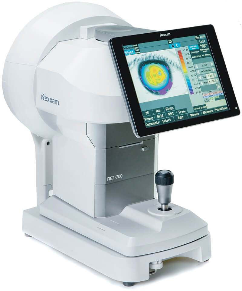
Rexxam RET 700
A Corneal Topography System is an advanced imaging technique that maps the surface of the cornea, the clear, front part of the eye. Similar to a 3D map that highlights terrain features, this system detects irregularities in the cornea’s curvature, which is typically smooth.
It aids doctors in identifying distortions, monitoring eye diseases, and planning surgical procedures. As an importer, wholesaler, and supplier of the Rexxam RET 700 Corneal Topography System in India, we provide a widely trusted solution for precise corneal mapping. Our compact, high-quality topography device is ideal for accurate eye diagnosis in ophthalmology clinics.
Corneal Topography System - Rexxam RET 700
All-in-One Model Featuring Auto Refractometer, Keratometer, Topographer, PC, and Database Auto Ref-Topographer Designed for Enhanced Functionality and Ease of Use

