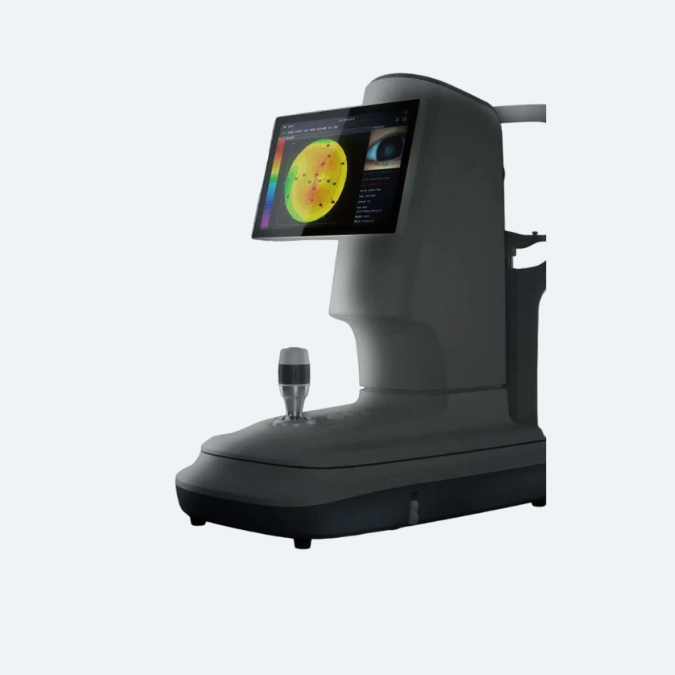
| Methodology | Spectral domain OCT |
| Scan wavelength | 840÷10 nm |
| Exposure power at pupil | ≤600 W |
| Working Distance | 34.9 mm
|
| Fixation | Both Internal as well as External |
| Scan speed | ≥86kA-scan/sec |
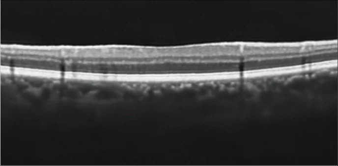
Posterior
SegmentScan | Scan depth | ≥3.5 mm |
| Axial resolution | <5um |
| Transverse resolution | ≤15um |
Types
Imaging
Options | Raster scan, single scan with adjustable orientation, dense cube scan, circle
or radial scan, 3D visualization, macular thickness map
|
Types
Analysis
Options | Retinal thickness map-RNFL thickness map with normative database for
glaucoma diagnosis, Optical nerve head analysis, optic disc scanning for
glaucoma, Progression analysis of RNEL, ONH or 2D, 3D modelling,
Enhanced depth imaging for choroidal layer scanning, Fovea to disc
alignment, auto disc centration or auto fovea finder, Posterior pole
symmetry analysis or combined ganglion cell+IPL and RNFL deviation
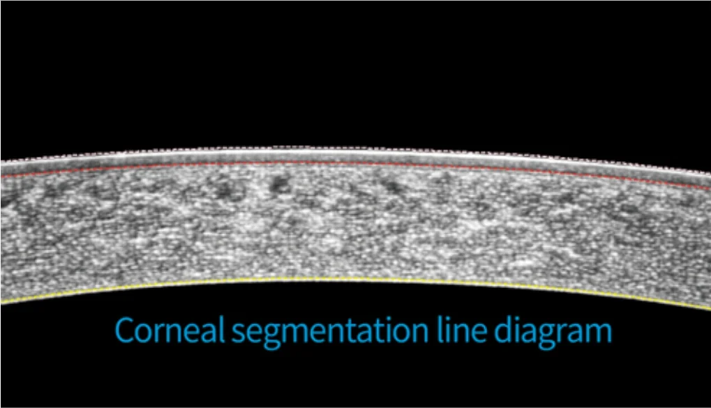
map for glaucoma diagnosis, Segmentation of different layer of retina
RPE elevation analysis or enface image analysis |
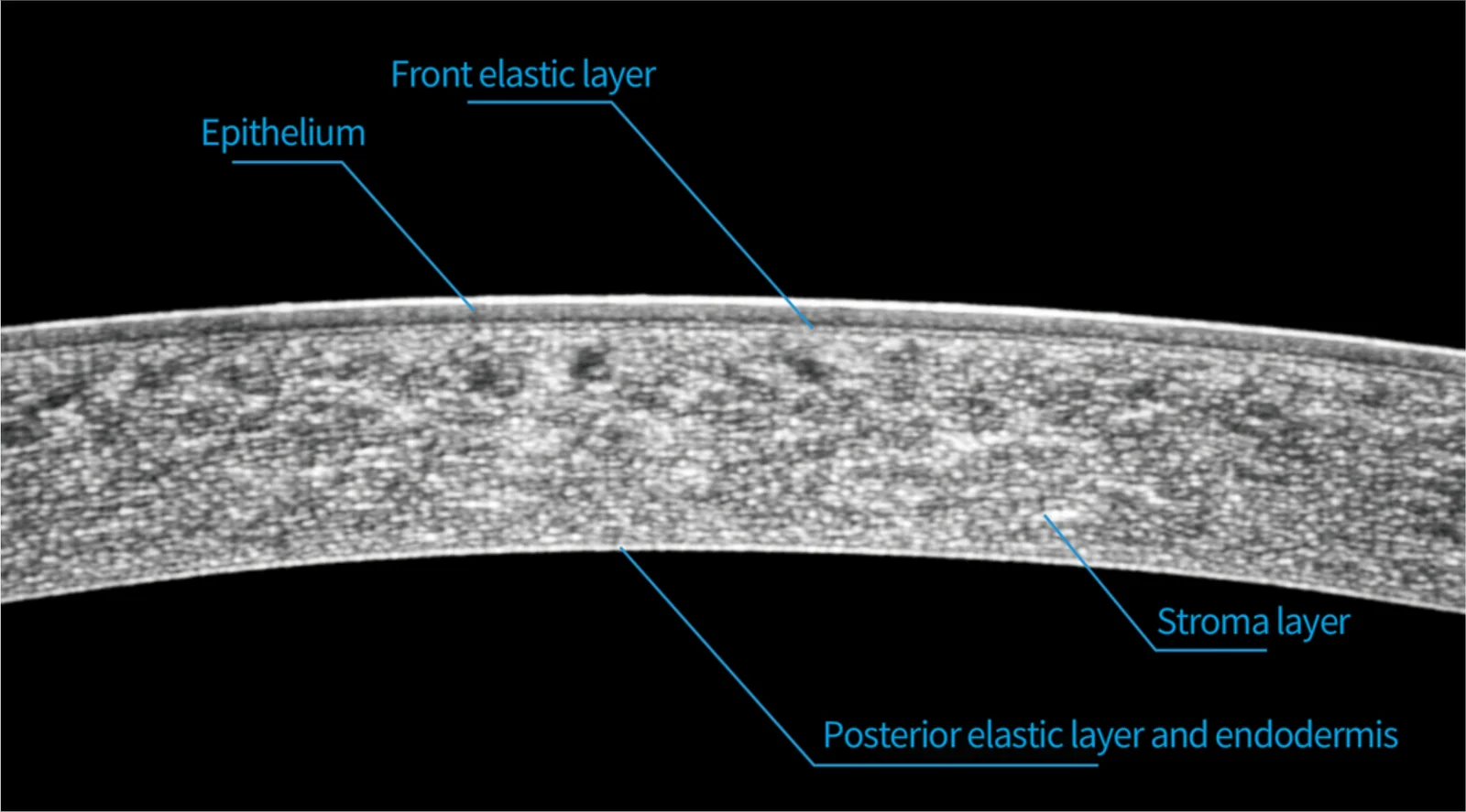
| Anterior Segment Scan | Scan depth | 23.5 mm |
| Axial resolution | <5um |
| Transverse resolution | ≤20um |
Anterior
Segment
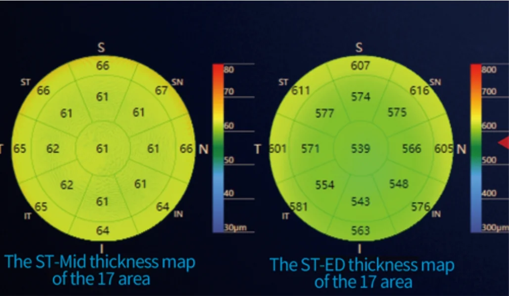
Imaging | Auto central corneal thickness (CCT), Anterior chamber angle view, Cornea
view
|
Accuracy
measurement | <3% |
| Type of Scan | Macular, Optic Disk, HD Scan |
No of A Scans
x B Scan | 512 A Scans x 128 B Scan, 200 A Scans x 200B Scans |
| A-Scan Depth | 13.5mm |
Center Wave
Length | 942 + 10nm |
| Light Source | Single SLD |
Type of
Imaging | Mono Color |
| Picture Angle | 45° x 30° |
Minimum
Photographable
Punil Diameter | 2.00mm |
| Depth Resolution | 3.5mm - 13.5mm
|
Vertical Scan
Range on
Fundus
| 13.5mm depth, Axial Resolution ≤ 5um |
Horizontal
Scan Range on
Fundus
| 13.5mm depth, Transversal Resolution ≤ 15um |
Vertical Scan
Range on
Cornea
| 3.5mm depth, Axial Resolution ≤ 5um |
Horizontal
Scan Range on
Cornea | 3.5mm depth, Transversal Resolution ≤ 20um |
| Lateral Resolution | Transversal Resolution ≤ 15um
|
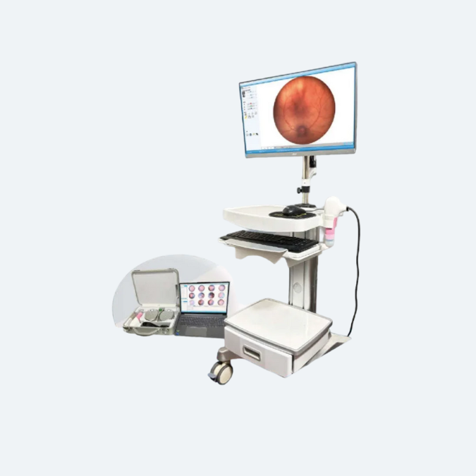
Fundus
imaging |
| Methodology | Line scanning
Ophthalmoscope (pSLO & IR ) |
Scan
wavelength | 942÷10 nm |
Exposure
power at
pupil
| ≤1500 uW |
| Field of view | Width: ≥45°
Height: ≥30°
|
| Frame rate | >7Hz |
Patient
interface | Internal fixation focus adjustment | -20D~+20D
|
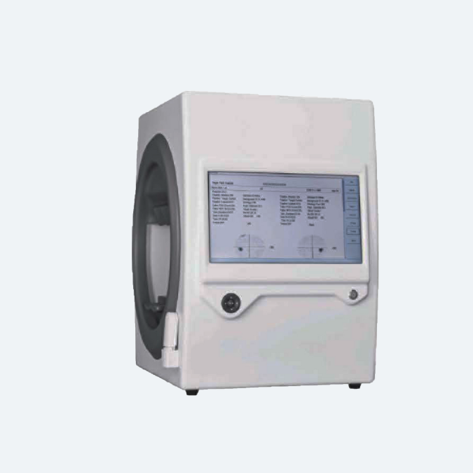
Physical
Specifications | Dimensions | 532Hx346Wx618D(mm)
|
| Weight | 35kg |
Software
Operating | CPU | i5 |
| Hard Disk | 1T or above |
| Conditions |
| Memory | 32G or above
|
| GPU | 8G or above
|
| Display resolution | 2560×1440 or above |
Operating
System
(OS) | Windows 10 and its compatible version |
| Operating Conditions |
| Input Voltage | 100-240V~
|
| Frequency | 50/60Hz
|
Input Power
| 100VA |
| Temperature | 10°C to +35°C
|
| Relative humidity | 30% to 90%
|
| Atmospheric pressure | 80 KPa to 106KPa |
Storage
Conditions |
| Temperature | - 10°C to +55°C
|
| Relative humidity | 10% to 95%
|
Atmospheric pressure
| 70 KPa to 106KPa
|
| Transport Conditions |
| Temperature | -40°C to +70°C
|
Relative humidity
| 10% to 95%
|
| Atmospheric pressure | 50KPa to 106Kpa
|
| Vibration, sinusoidal | 10Hz to 500 Hz:0,5g
|
| Shock | 30g, duration 6ms
|
| Bump | 10g, duration 6ms
|