Fundus Camera - EFC3
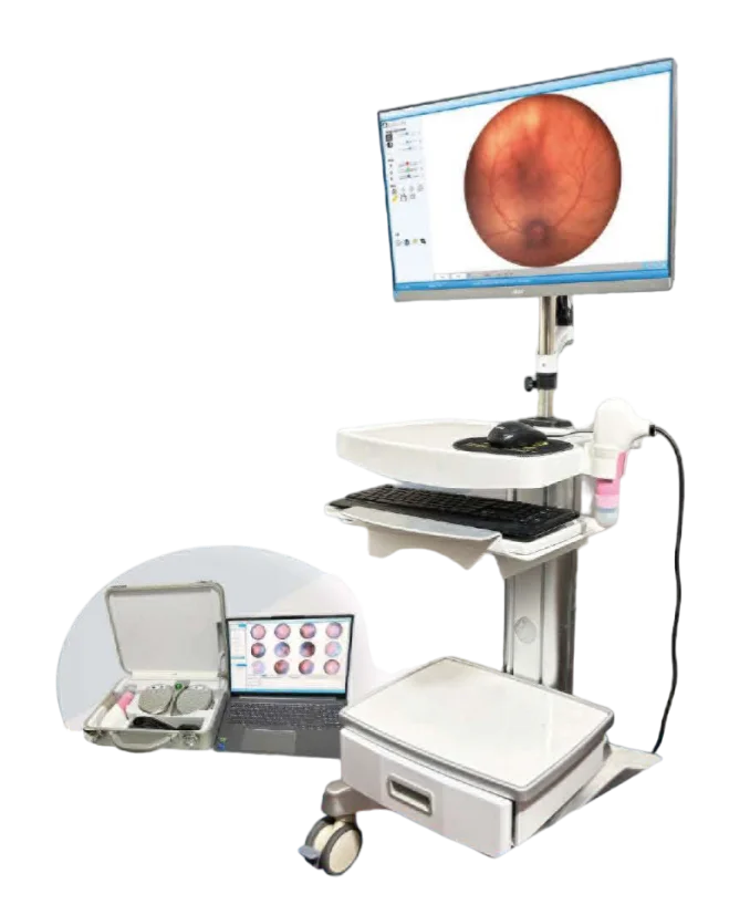
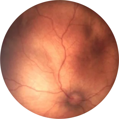
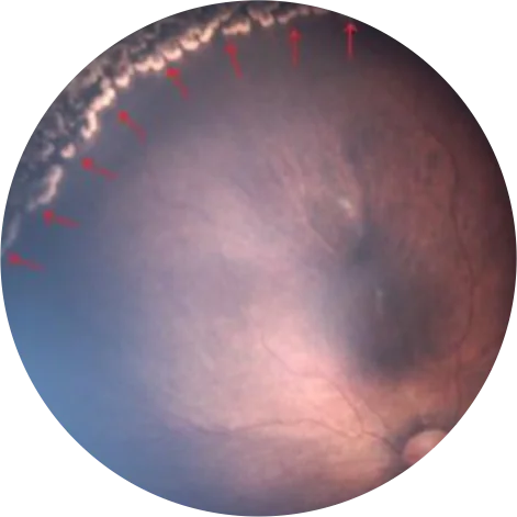
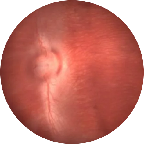
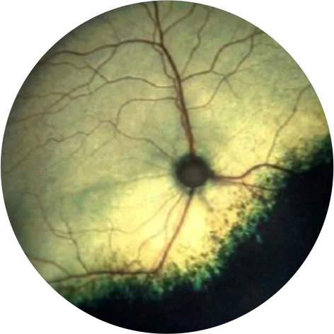
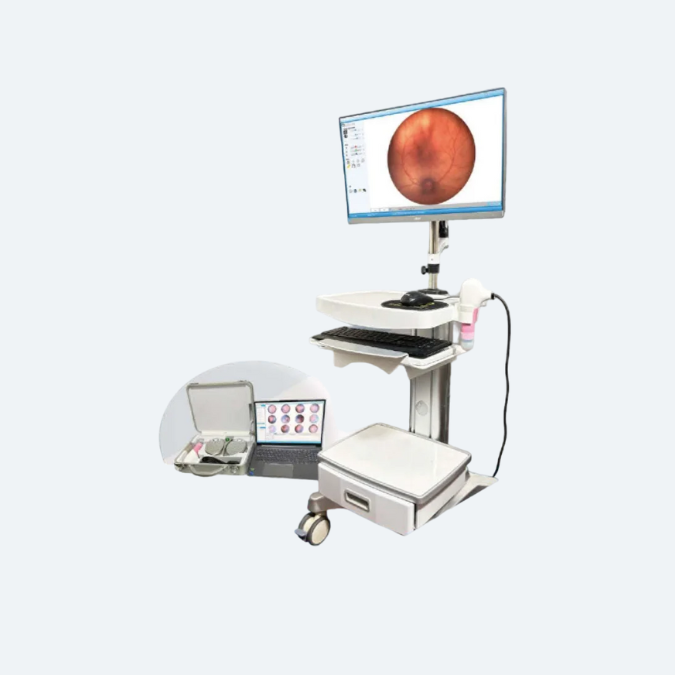
The EFC3 Wide Field Retina Camera is a high-performance ophthalmic imaging device designed for capturing detailed wide-angle images of the retina. It features a wide field of view, allowing clinicians to visualize a larger portion of the retina in a single image.
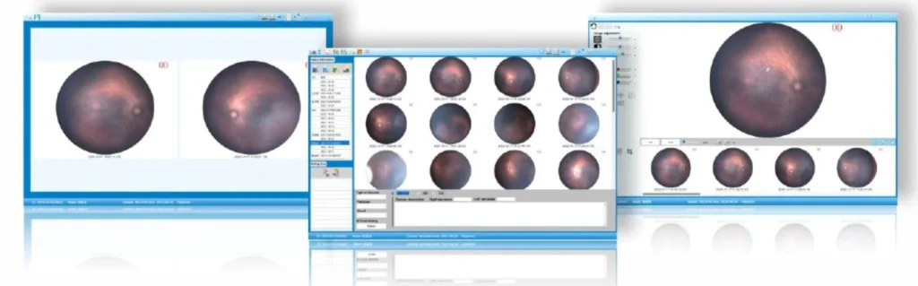
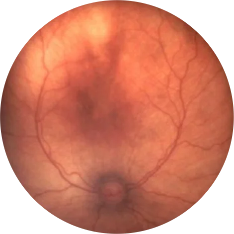
The camera’s advanced optics and imaging technology provide high-resolution, color-accurate images, essential for diagnosing and monitoring various retinal diseases, such as diabetic retinopathy, macular degeneration, and retinal detachment.
Key features of the EFC3 camera may include:
- Wide field of view: Typically capturing up to 133° of the retina in one shot, allowing comprehensive examination with fewer images.
- High-resolution imaging: Ensures clear and detailed visualization of retinal structures.
- Non-mydriatic imaging: Captures retinal images without the need for pupil dilation, offering a more comfortable experience for patients.
- Ease of use: Designed with user-friendly software and ergonomic controls to simplify the imaging process.
- Versatility: Suitable for both routine screenings and detailed diagnostics in ophthalmic practices.
System Introduction
- The system is easy and quick to use
- Image history to view the progress of the disease
- Images can be freely labelled
Multiple Use Mode
- Trolley mode
- Portable mode, laptop direct connection for easy operation, can be bedside and ward
Extra large filed view
Large 133° field view for more fundus information in a single shot
133º Field View Diagram
ROP lesion area
Laser Funduscopy
Real Photograph
Anima Ifilming System
Can be used directly in animal clinical trials
Rabbit Fundus
Cut Funduscopy
Technical Specification EFC3 Fundus Camera
Item | EFC3 |
Visual field range | 133° |
Minimum pupil | 4mm |
Collector | Medical-grade professional CMOS |
Pixel | >=800 million |
Light source adjustment | Seamless adjustment light |
Lighting Method | Built-in integrated lighting |
Shooting Method | Foot switch (manual) / software (auto) shooting |
Focus mode | Auto focus |
Lesion marking | Auto marking |
Shooting mode | Colour photography + Fluorography |
Port | USB, DICOM |