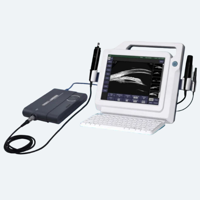
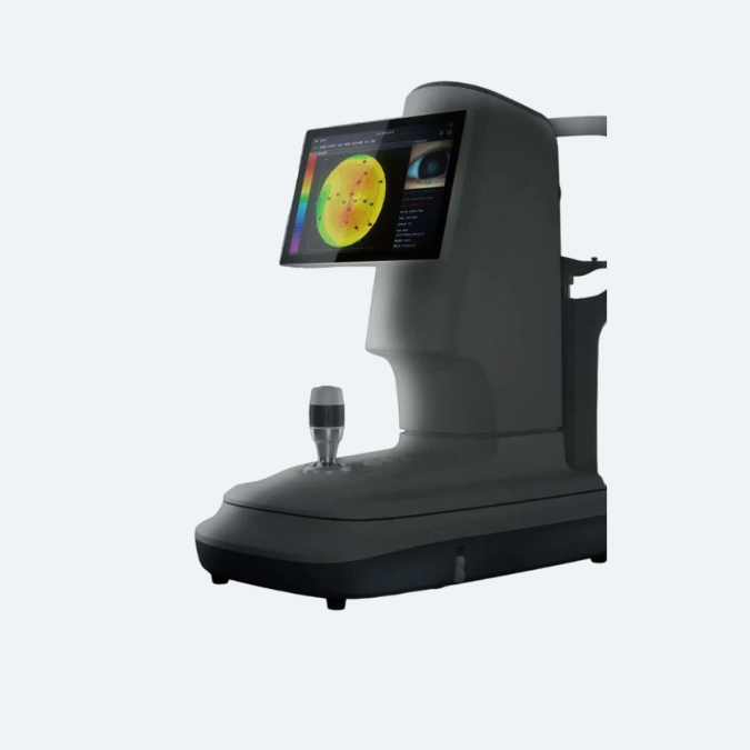
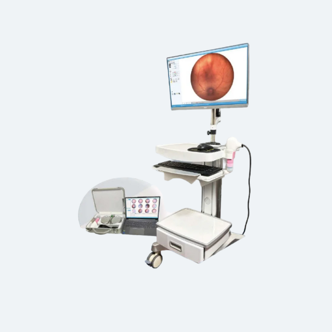
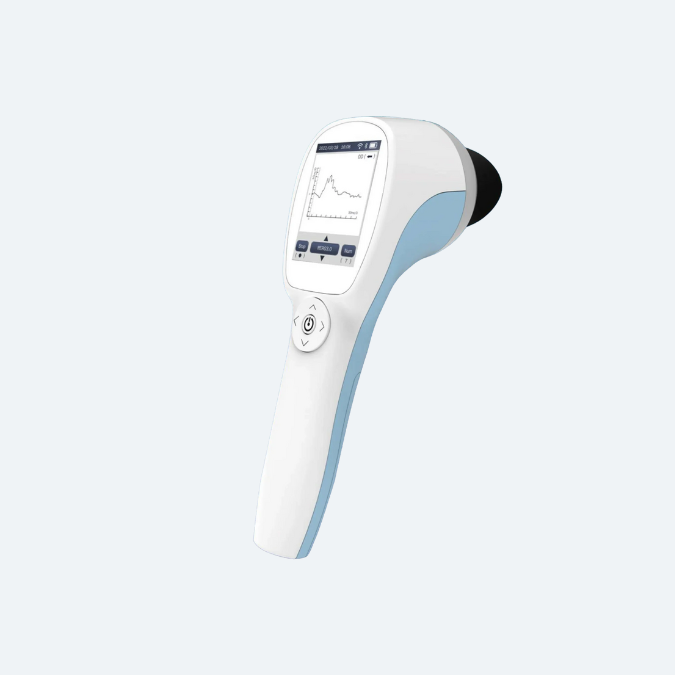
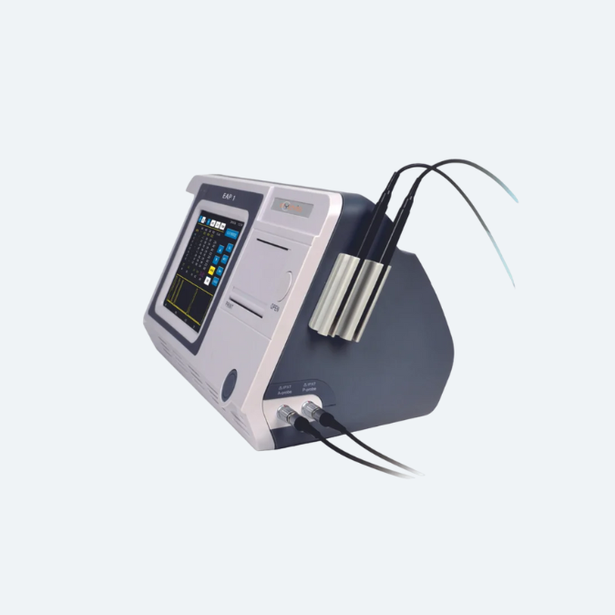
Ophthalmic Ultrasound A-Scan/Pachymeter System - EAP 1
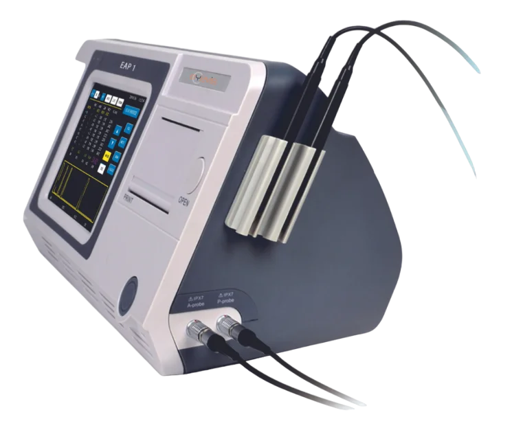
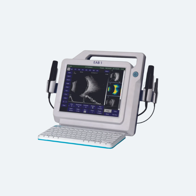
The EAP 1 Ultrasonic Biometer is an essential ophthalmic tool for accurately measuring the eye’s axial length, a critical step in determining intraocular lens (IOL) power prior to cataract surgery. Utilizing ultrasound technology, it delivers precise measurements of key internal structures such as the cornea, lens, and retina. This ensures enhanced precision in both refractive and cataract surgeries. The device is non-invasive, offering fast and reliable results for improved surgical outcomes.
Ultrasound
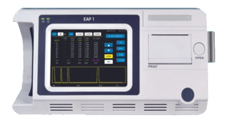
A-Biometer
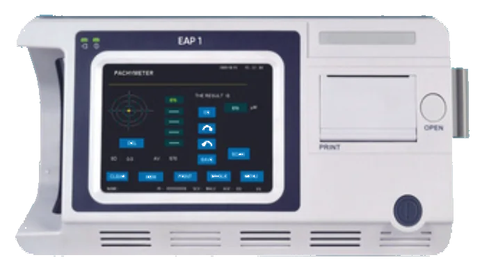
Pachymeter

A-Biometer / Pachymeter
Accessories
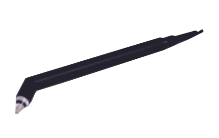
20MHz straight P-probe
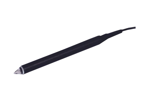
20MHz angled P-probe
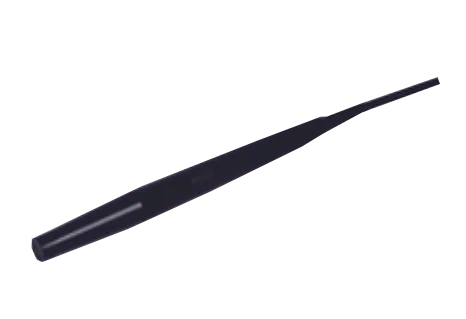
10MHz A-probe
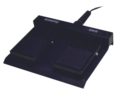
Footswitch
Pachymetry
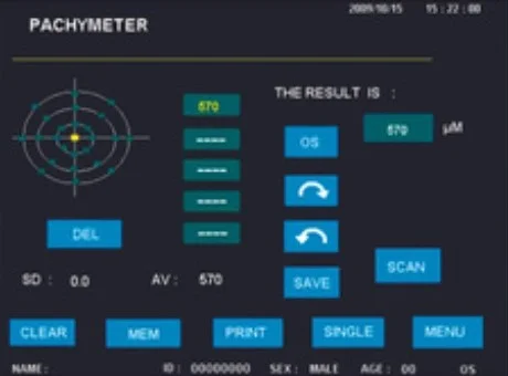
Accurate Results
Automatic reading at single or multiple points for corneal thickness; Multiple measurements at single point for higher reliability; Higher accuracy enabled by averaging readings.
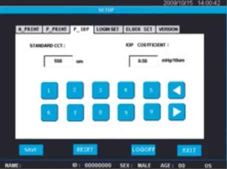
IOP Adjustment
Intraocular pressure adjustment provides reference for tonometer measurement; Parameter adjustability based on user’s experience.
Management
Patient Management
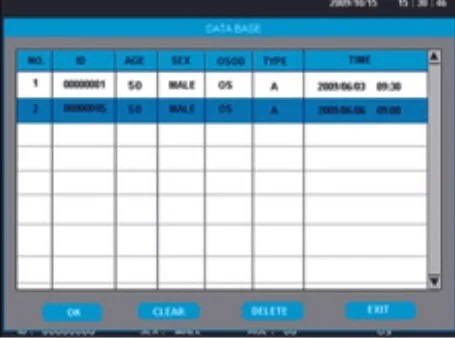
Includes a built-in data archiving feature with capacity to store up to 180 patient records for easy access and management.
User-defined Preference
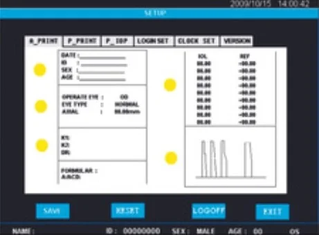
Users can customise acoustic velocities, set IOP parameters, and configure printing options to suit their specific requirements.
Instant Printout
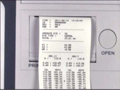
One-click printout with the built-in thermal printer, featuring user-defined print settings for customised reports.
A-Biometry
Precise & Accurate
MEDA’s advanced technology and expertise in ophthalmology ensure A-Scan precision and accuracy in both cataract and immersion modes for reliable results.
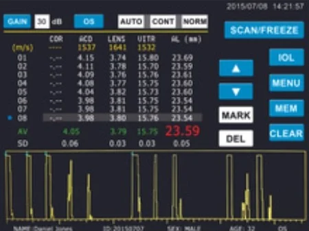
Reliable
Automatically measures up to 8 groups of readings per session, with averaging and standard deviation calculations for enhanced accuracy and reliability.
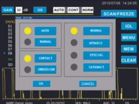
Comprehensive
Equipped with a touch screen and footswitch for seamless and efficient operation.
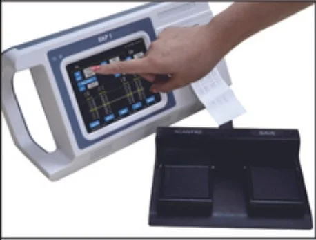
Convenient
Users can customise acoustic velocities, set IOP parameters, and configure printing options to suit their specific requirements.
IOL
IOL Formulae
Includes 6 widely used IOL calculation formulae and 5 key formulae for post-refractive IOL calculations, ensuring accurate lens selection.
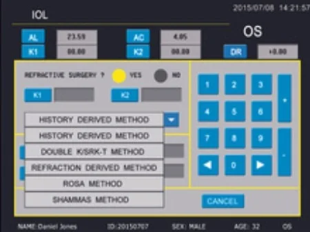
Intuitive Interface
Allows instant switching between different formulae and features a dual-formula display for easy and direct result comparison.
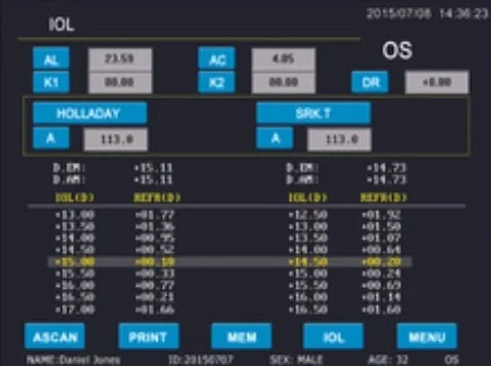
Simple Operations
Offers enhanced database accessibility with a single-click instant printout for quick and efficient reporting.
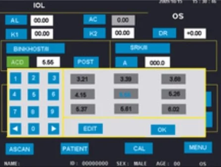
Tight Integration
Provides seamless switching between A-scan and IOL calculations, with automatic import of axial length from A-scan measurements for faster workflow.
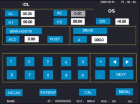
EAP 1 A scan with Pachymeter Technical Specifiaction
| Specification | Details |
|---|---|
| Probe | 10MHz with Fixation Red Light |
| Total Gain | 100dB with an adjustable range of 0–50dB |
| Biometry Accuracy | ±0.05mm |
| Resolution | 0.01mm |
| Measuring Range | 15–40mm |
| Measuring Mode | Contact or Immersion |
| Measuring Parameters | Anterior Chamber Depth, Lens Thickness, Vitreous Length, and Axial Length |
| Measuring Modes | Automatic (Normal, Cataract, Aphakic, and Special) and Manual |
| Groups of Readings | 8 Groups of Readings with Averaging and Standard Deviation |
| Standard Configuration | |
| Standard Configuration | 10MHz Probe, 20MHz Probe, Footswitch, Test Object, AC Adapter |
| IOL Calculation | |
| General | SRK-II, BINK-II, HOFFER-Q, History-derived, Refraction-derived, SHAMMAS |
| Post-Refractive | SRK-T, HOLLADAY, HAIGIS, Double K/SRK-T, ROSA |
| Pachymeter | |
| Probe Frequency | 15–20MHz |
| Display Resolution | 1μm |
| Biometry Accuracy | ±5μm |
| Measuring Scope | 230–1200μm |
| Maps | Multiple Corneal Maps with Graphical Display |
| General | |
| Power Supply | AC 100–240V, 50/60Hz, 50VA |
| Dimensions | 337mm x 177mm x 155mm (L x W x H) |
| Weight | 1.7Kg |
| Optional | |
| Optional | Immersion Shell |

