Corneal Topography System & Imaging
Corneal topography System is a special photography technique that maps the surface of the clear, front window of the eye (the cornea). It works much like a 3D (three-dimensional) map of the world, that helps identify features like mountains and valleys. But with a topography scan, a doctor can find distortions in the curvature of the cornea, which is normally smooth.
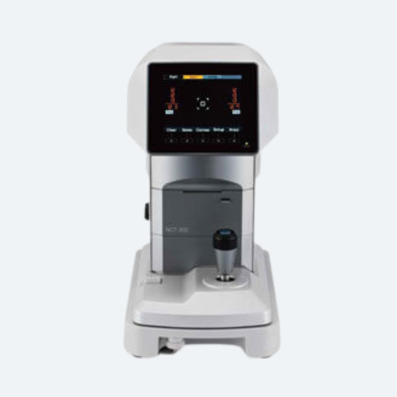
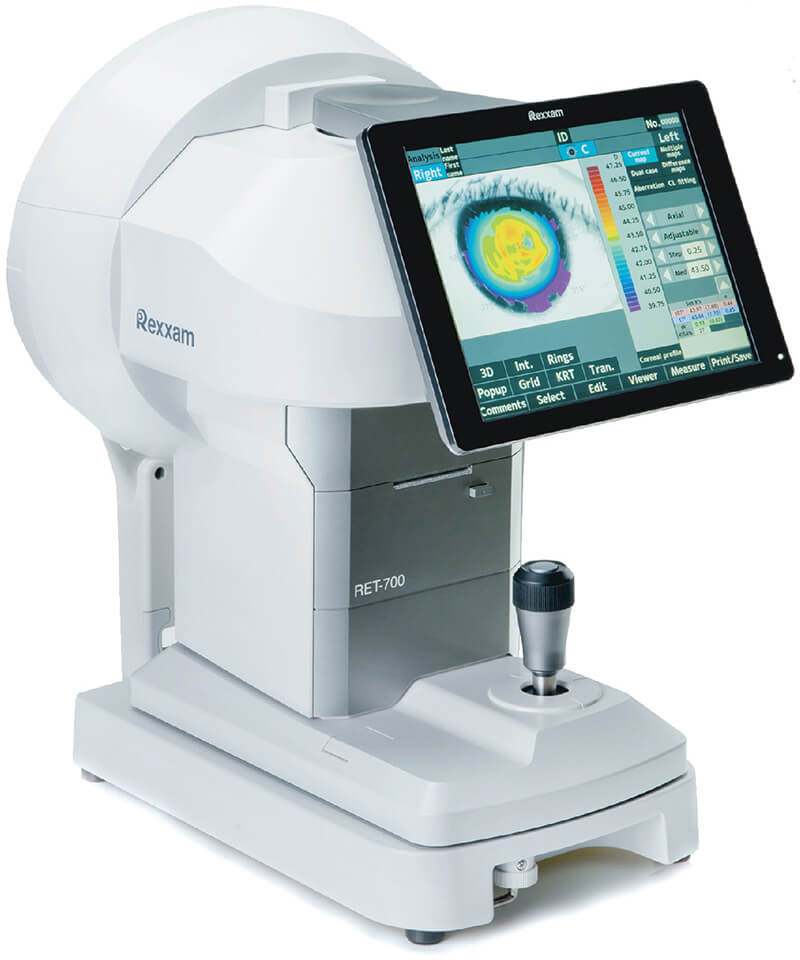
It also helps doctors monitor eye disease and plan for surgery. We are Importer, Wholesaler & Supplier of Rexxam RET 700 Corneal Topography System in India. The widely used Corneal Topography System helps to map the surface of the clear, front window of the eye. We offer finest, compact corneal topography device suitable for eye diagnosis at any ophthalmology clinics.
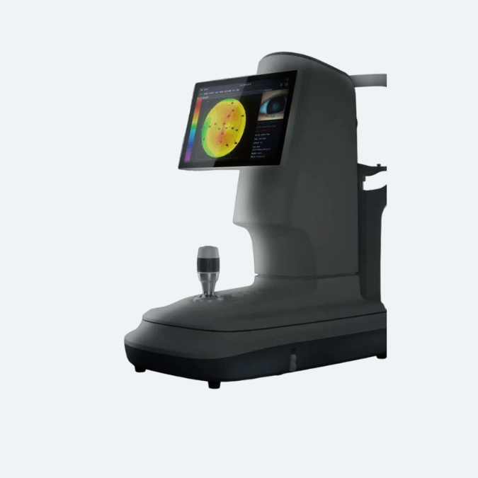
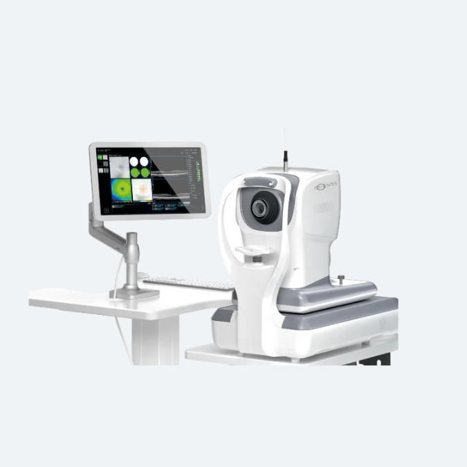
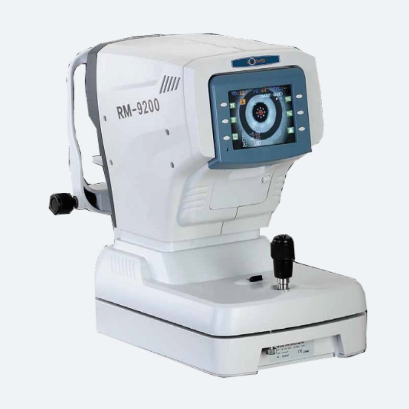
Corneal Topography System - Rexxam RET 700
All-in-one model including auto ref, keratometer, topographer, PC and database
Auto Ref-Topographer with pursued functionality and operability
Corneal Topography System Overview
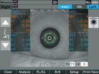
All-in-one
Measurements of the auto ref, kerato and topography are taken at the same time. Maximum 6 images of topography are captured continuously.
Wide Topo Measurement Range
The measurement range is from 0.4mm to 10.7mm(R8.0).
Also, the peripheral corneal (approx. 16.00mm) is measurable.
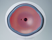
A Variety of Analysis Function
A variety of analysis display includes Current map, Multiple map, Dual case, Difference map, Aberration and CL fitting etc.
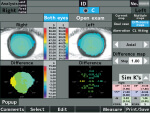
Difference map
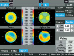
Abberation
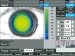
CL fitting
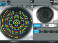
Ring Edit Function
A ring can be assigned manually if the ring cannot be measured automatically.
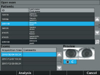
Database
Measurement data can be stored and accessible any time.
Scotopic & Photopic Pupil Diameter Measurement
Both scotopic and photopic measurements are available.
※ S.P.S: Scotopic Pupil Size
P.P.S: Photopic Pupil Size
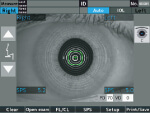
Scotopic measurement
[S.P.S. function]
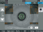
Photopic measurement
[P.P.S. function]
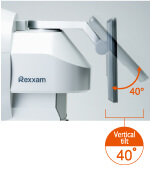
Wide Screen
10.4 inch wide color screen.
The swivel/tilt function allows the operator to support easily the patient during operation.
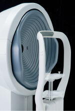
Electric Chinrest
It is easy to align the eye position of the patient with the eye mark.
Corneal Topography System Specification
| Function | |
| Eye refraction measurement | |
| Spherical power | -20D to +30D (step:0.12D/0.25D) VD=0 |
| Cylindrical power | 0D to ±10D(step:0.12D/0.25D) |
| Axis angle | 1 to 180 degrees (step:1°/5°) |
| Measurement range of cornealcurvature | φ2.0mm |
| Corneal curvature radius | |
| Corneal curvature radius | 4.90mm to 10.10mm (step:0.01mm) |
| Corneal refractive power | 68.88D to 33.42D(step:0.12D/0.25D) *corneal refractivity = 1.3375 |
| Corneal astigmatism | 0D to ±10D(step:0.12D/0.25D) |
| Axis angle | 1 to 180 degrees (step:1°/5°) |
| Measurement of corneal shape | |
| Measurement range | φ0.4 – φ10.7(R8)mm |
| Pattern of measurement light | 19 cocentric circle ring patterns |
| Measurement point | 6,200 |
| Working distance | 77.5mm |
| Peripheral cornea | Approx.φ16(R8)mm |
| Axial | – |
| Tangential | – |
| Elevation | – |
| Refractive | – |
| Zernike | – |
| Fourier | – |
| Type | Placid Dome |
| Fitting of contact lens | – |
| Dry eye observation function | – |
| Meibomian observation function | – |
| Measurement of pupil diameter | φ2.0mm to φ8.5mm(step:0.1mm) |
| Alignment method | Manual alignment |
| PC | Built-in |
| Monitor | 10.4 inches touch panel colored LCD (XGA) |
| Printer | Thermal line printer (paper width 58mm) |
| External interface | Ethernet(10/100Mbps)×1, USB-A×2, USB-B×1 |
| Source voltage/frequency | AC 100 to 240V, 50/60Hz |
| Power consumption | 90VA |
| Power saving function | OFF,3,5,10min. (switchable) |
| Size | H(507mm)×W(346mm)×D(422mm) |
| Weight | 17kg |
| Standard Accessories | Operation manual, Power cord, Printer paper, Fuse, Dust cover, Model eye, Chinrest paper pin |
Corneal Topography System & Imaging : What To Expect & How To Interpret Results?
What Is Corneal Topography?
Corneal topography is an advanced imaging technique used to map the surface of the eye’s clear, front layer, known as the cornea. Similar to how a 3D map highlights geographical features like mountains and valleys, this scan reveals irregularities in the cornea’s curvature, which is typically smooth. This detailed mapping assists doctors in detecting distortions, monitoring eye diseases, and planning surgical procedures.
What Conditions Is Corneal Topography Used for?
Scarring: Injuries or infections can scar the cornea, altering its shape. A topography scan accurately measures this distortion and its impact on vision.
Growths: Topography allows for the monitoring of pterygia or other growths on the cornea by tracking their size.
Astigmatism and Keratoconus: This scan aids in the detection of astigmatism and early-stage keratoconus, as well as in monitoring their progression.
Contact Lens Fitting: Topography helps determine the most suitable type of contact lens for improving vision. In cases of significant distortion, a specialized hard contact lens (RGP) may be recommended to correct vision.
Corneal topography can be utilized for a wide range of purposes.
- Planning and Monitoring Refractive Surgery
- Post-Refractive Surgery Ocular Health Monitoring
- Determining Suitable Intraocular Lens for Cataract Surgery
- Evaluating and Treating Astigmatism After Keratoplasty
- Detection of Corneal Conditions: Including pterygia, corneal scars, and Salzmann nodules.
- Ocular Disease Monitoring
- Assessing Anterior Angle Integrity for Glaucoma Patients
- Measuring Corneal Depth
- Evaluating Corneal Nerve Health
- Detection of Infectious Keratitis
- Examination of Corneal Endothelium
How Does Corneal Topography Help with Surgery?
Refractive surgery: During refractive surgery like LASIK, the shape of the cornea is changed to correct refractive errors like myopia (nearsightedness). A topography scan helps the surgeon understand how to precisely reshape the cornea.
Cataracts: When cataracts make the eye’s natural lens cloudy, it is replaced with an intraocular lens (IOL) during cataract surgery. Corneal topography helps surgeons select the right IOL in some cases.
Corneal transplants: After a corneal transplant, a surgeon may use corneal topography to help a patient heal correctly. The images help assess which stitches should be removed and when based on the shape of the cornea.
Corneal cross-linking: Corneal cross-linking surgery helps strengthen a cornea with keratoconus. A topography scan may be done to see if this surgery is needed. After surgery, scans are done to monitor the eye.
What to Expect During a Corneal Topography Scan?
- You will be seated facing a large bowl with lighted circles inside it.
- The chin and forehead rests keep your head secure to get the clearest images.
- You will be asked to stare at a fixed target in the bowl while the pictures are taken.
- The scan only takes a few seconds, but it may need to be repeated a few times.
- Getting a corneal topography is painless, as nothing touches your eye during the scan.
Types of Corneal Topography
There are three different types of technologies used for corneal topography:
Placido Disc Reflection Systems: These systems measure the curvature, irregularities, tear film quality, presence of foreign bodies, and other aspects of the anterior cornea. The accuracy of these measurements depends heavily on the tear film, which reflects light.
There are two types: small-cone and large-cone systems. Small-cone systems are more precise as they gather more data points, while large-cone systems are easier to operate and simplify the data collection process.
Scheimpflug and Scanning-Slit Topography: These two methods provide detailed information about both the anterior and posterior cornea. They are particularly useful in detecting and managing corneal swelling, which is crucial for contact lens wearers.

