Ultrasonic A/B Scanner with UBM
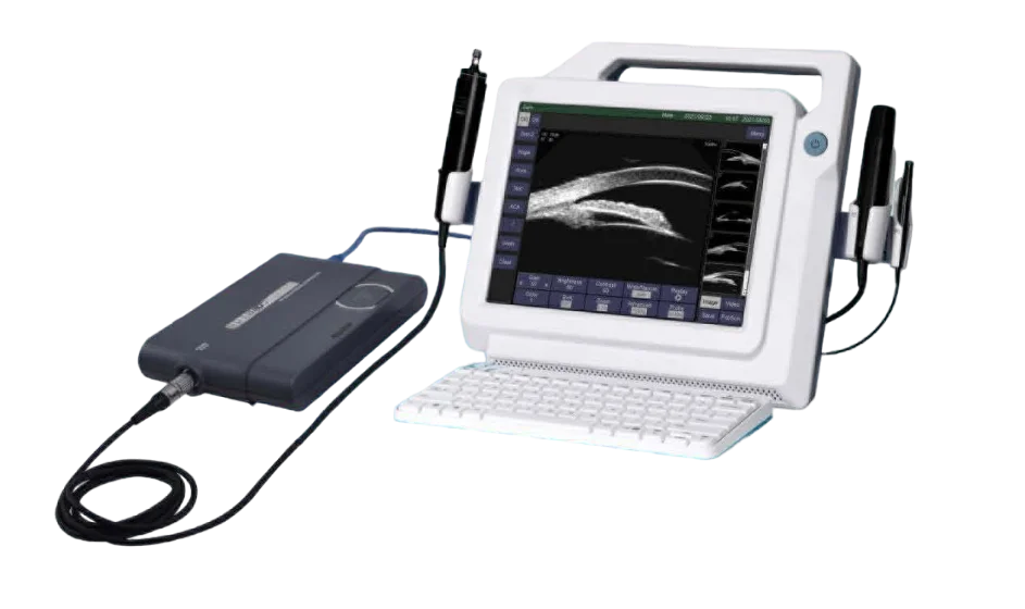
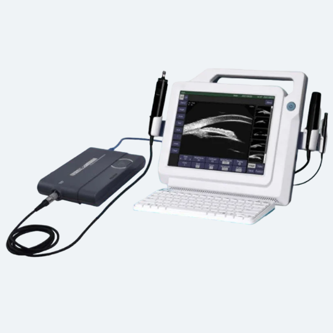
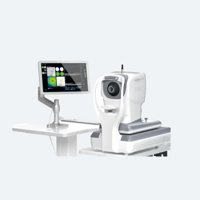
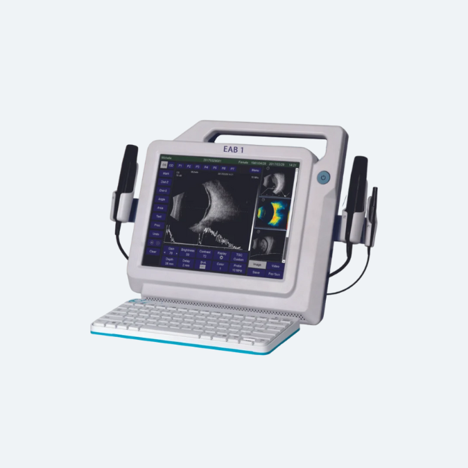
The EUB-1 Ultrasonic A/B Scanner with UBM is an advanced diagnostic tool designed for comprehensive ophthalmic imaging. Combining A-scan, B-scan, and Ultrasound Biomicroscopy (UBM) capabilities, this versatile device delivers high-resolution imaging of both anterior and posterior segments of the eye.
Its compact and portable design makes it suitable for various clinical settings, while the intuitive interface ensures ease of use. With precise measurement tools and enhanced imaging clarity, the EUB-1 is an essential solution for accurate diagnosis and management of complex eye conditions.
Professional Ultrasound
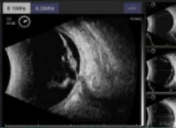
B-Scan Enhanced Visibility and User-friendliness
- Image/Video Buffer Slots: Instantly capture, review, and compare images or videos with ease.
- Real-Time Capture: Images and videos are recorded in real-time and stored in lossless formats (.bmp and .avi).
- Enhanced Observation: Features zoom and adjustable scan depth for detailed analysis of ocular diseases.
- Vitreous-Enhanced Mode: Improves visibility of the vitreous body for more accurate evaluations.
A-Scan & IOL Calculation Enhancements and Tight Integration
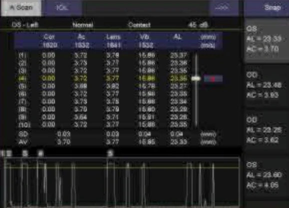
- Biometry Buffer Slots: Instantly capture, review, and compare biometric data with ease.
- Enhanced Accuracy: Achieves greater precision with Average and Standard Deviation calculations for up to 10 scans per exam.
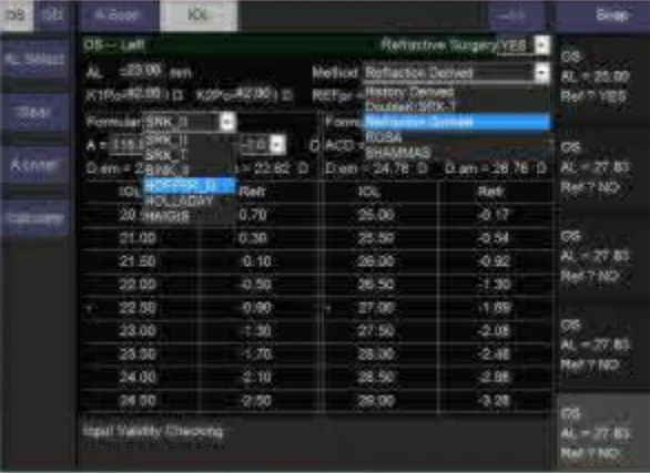
- Comprehensive IOL Options: Offers 6 commonly used IOL formulae and 5 post-refractive IOL formulae for versatile lens calculation.
Full Reporting Capabilities
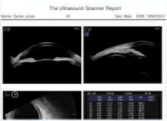
Comprehensive report combining A-scan/IOL results, B-scan images, and doctor’s notes, featuring a customizable dictionary for symptom entries.
Comprehensive Data Archiving and Management
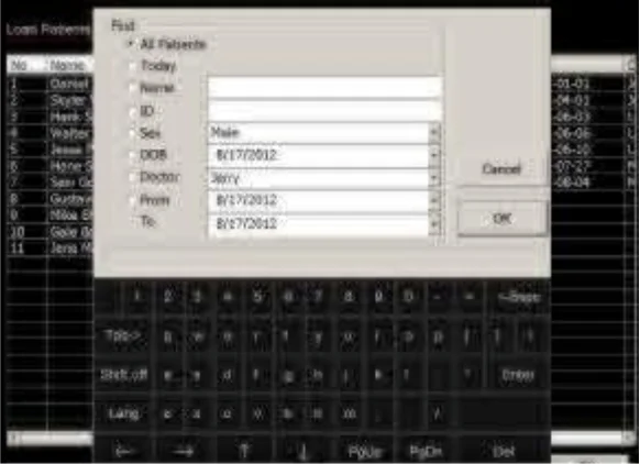
The simple user interface enables easy loading, searching, printing, and more. Offers unlimited storage capacity for over 20,000 exams, each containing up to 8 lossless images.
Features
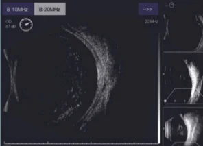
- High-Definition Imaging: Features a 50MHz UBM and 10MHz B-scan for superior image clarity.
- Touchscreen Display: Equipped with a high-resolution LCD touchscreen for intuitive operation.
- Real-Time Image/Video Storage: Capture and store images/videos in real-time across multiple buffer slots for instant comparison and review.
- Customizable B-Scan Options: Offers multiple TGC settings, including a vitreous-enhanced mode, to suit operator preferences.
- Dual UBM Fields of View: Provides full or half sulcus-to-sulcus images with two fields of view (16mm × 11.5mm and 8mm × 5.7mm).
- Comprehensive Clinical Reporting: Editable reports integrate UBM/B-scan images, A-scan/IOL results, and customizable textual comments.
- PDF Reports: Generate reports in PDF format for easy sharing and printing.
- Printer Compatibility: Supports both graphical and textual printers.
- Versatile Connectivity: Includes HDMI and USB 2.0 ports for flexible connections.
- HDMI Output: Enables dual-screen display for enhanced showcasing and collaboration.
Technical Specification
| B-Scan & UBM | |
|---|---|
| Ultrasound Probes |
|
| Axial Resolution |
|
| Lateral Resolution |
|
| Scan Depth |
|
| Cineloop | 10s/100 frames with dynamic replay |
| Image Acquisition |
|
| Gray Scale | 256 Levels |
| Gain |
|
| TGC (for B-Scan) |
|
| Color Codes | 8 |
| Measurements |
|
| Biometrics A-Scan | |
| Probe | 10MHz with Fixation Red Light |
| Gain | 1-60dB |
| Measuring Method | Contact or Immersion |
| Measuring Range | AL Range: 15mm-40mm |
| Measuring Accuracy | ±0.05mm |
| Measuring Mode | Automatic (Normal, Aphakic, Special and Cataract) or Manual |
| Measurements | Average and Standard Deviation for up to 10 scans per exam Configurable Tissue Velocities under Special or Manual mode |
| IOL Calculation | |
| General |
|
| Post-Refractive |
|
| General | |
| Display | High-Resolution 12.1" LCD |
| Printer Compatibility | Graph/Text Printer and Video Printer (PAL) |
| Interface | Video-out (PAL), HDMI, USB 2.0 Ports |
| HDD | 500GB or higher |
| Network | Folder/Report Sharing |
| Power Supply | AC 100-240V, 50/60 Hz |
| Operations | Touch Screen Wireless Mouse & Keyboard Footswitch |
| Optional | Eye Cup Immersion Shell Video Thermal Printer |

