Ocular Diagnostic Master - ETD1
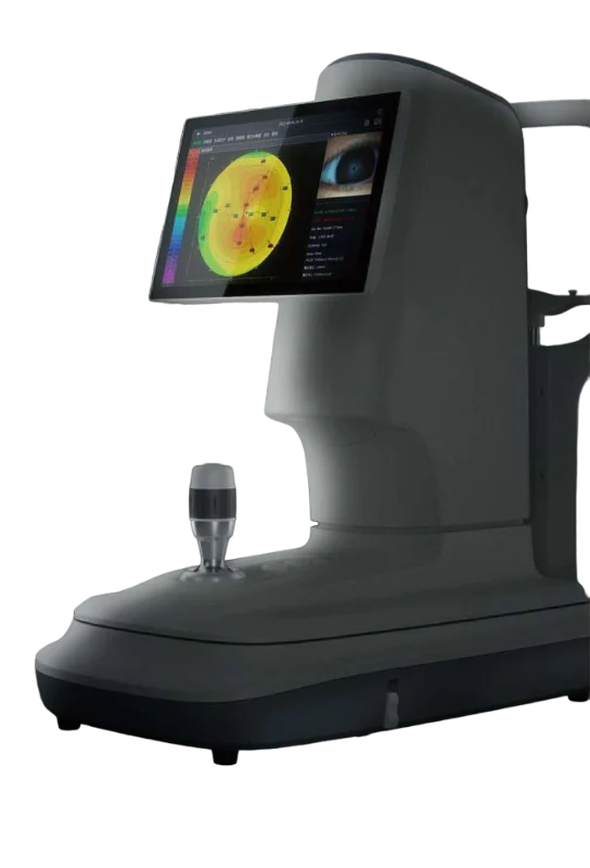
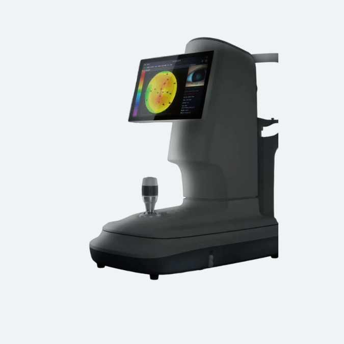
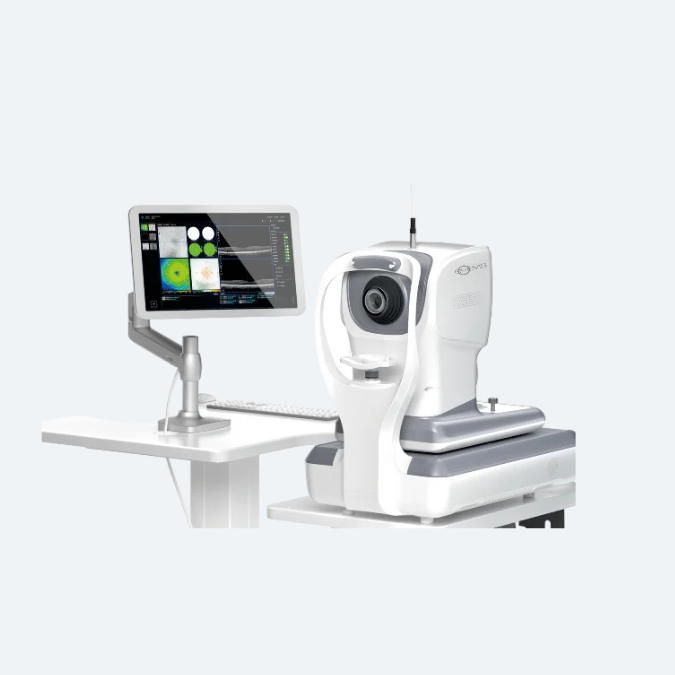
The ETD1 is a cutting-edge, multi-purpose corneal topographer that integrates corneal topography analysis with dry eye assessment. Designed for comprehensive ocular evaluations, it provides precise measurements to detect irregularities in corneal shape and structure while also identifying signs of dry eye syndrome.
This dual-function device is essential for improving diagnostic accuracy in various eye conditions, enhancing patient care. Suitable for clinics and research environments, the ETD1 ensures efficient and reliable assessments for both corneal health and tear film stability.
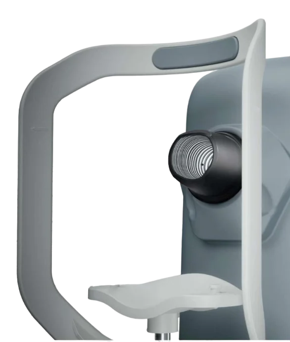
Placido Ring
- Thousands of measure points – ensure more data available and accurate analysis
- Smaller cone design – bigger projection area
- 3 Illuminations – white illumination, infrared illumination, cobalt blue illumination
ETD1 9 Functions
| Dry Eye Diagnosis | Topography |
|---|---|
| Non-Invasive Tear Film Breakup Time | Topography Analysis |
| Cornea Sodium Fluorescein Staining | Pupil & Corneal Diameter Measurement |
| Non-Invasive Tear Meniscus Height | |
| Eyelid Margin | |
| Meibomian Glands Function Evaluation | |
| Conjunctival Redness Analysis | |
| Lipid Layer Thickness |

ETD1 9 Functions
- Integration design enables maximum treatment room utilization
- Dry eye diagnosis and Topography analysis integrated
- 10.1″touchscreen, ease of operation
Doctor-Patient Communication
- Integration design enables maximum treatment room utilization
- Dry eye diagnosis and Topography analysis integrated
- 10.1″touchscreen, ease of operation
Ergonomic Design
- Integration design enables maximum treatment room utilization
- Dry eye diagnosis and Topography analysis integrated
- 10.1″touchscreen, ease of operation
ETD1 9 Functions
- Integration design enables maximum treatment room utilization
- Dry eye diagnosis and Topography analysis integrated
- 10.1″touchscreen, ease of operation
Dry Eye Diagnosis - Ocular Diagnostic Master
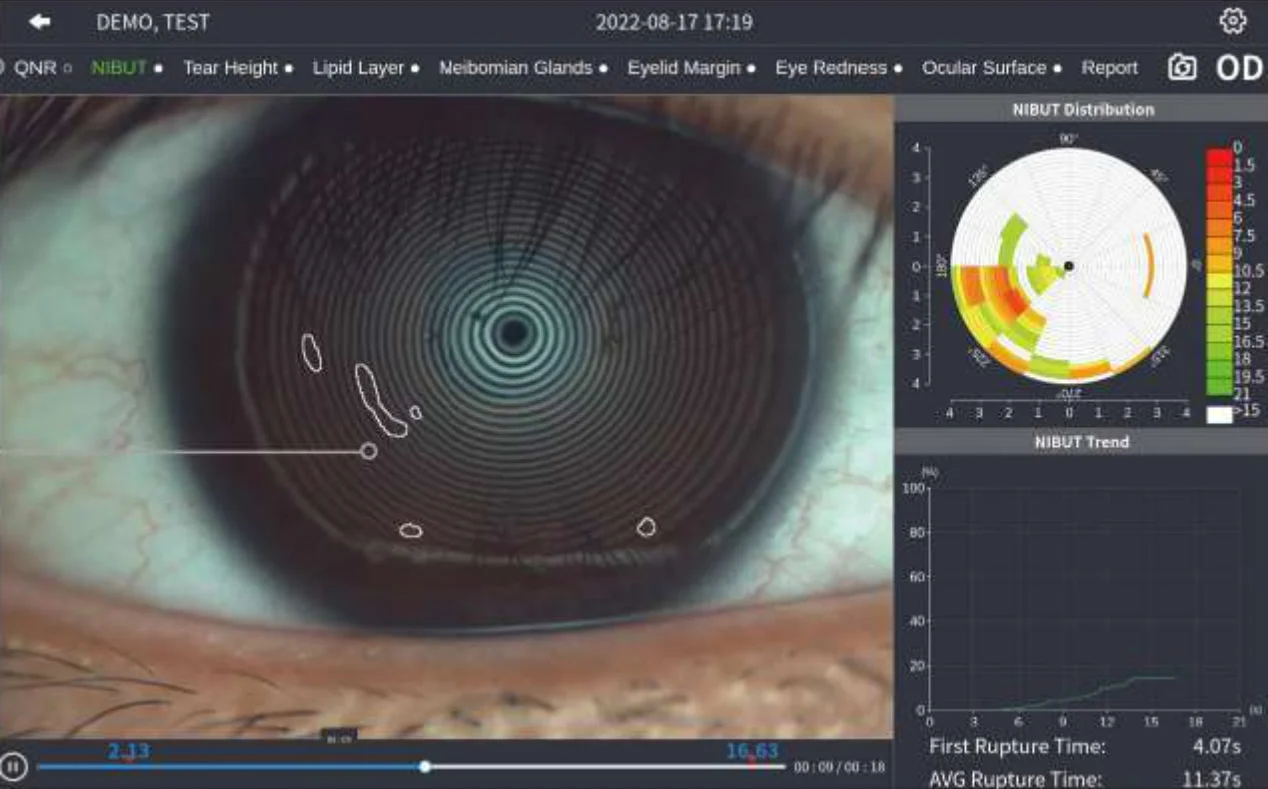
Non-Invasive Breakup Time
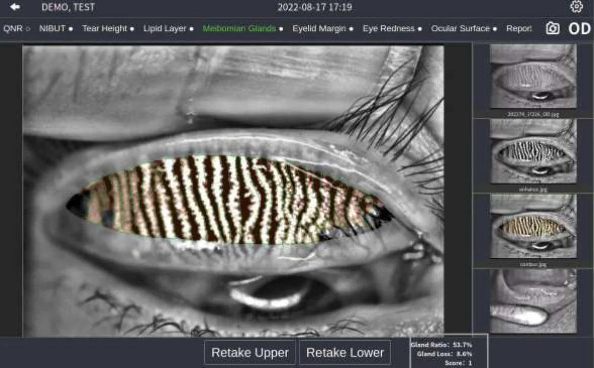
Meibomian Glands Function Evaluation
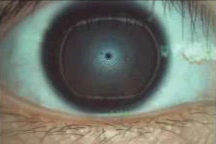

Automatic identification system depicts tear meniscus area and measures the tear height intelligently.
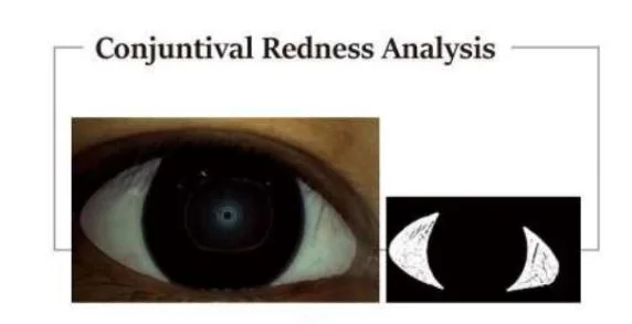
Identify and calculate percentages of conjunctival congestion and ciliary congestions and evaluate severity of eye congestion
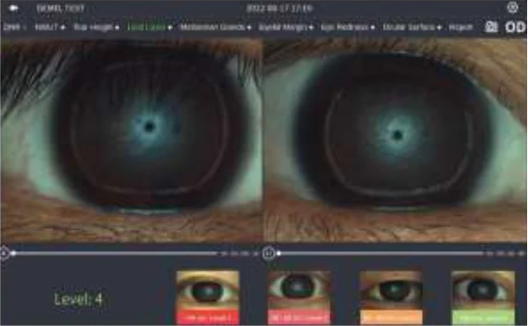
Lipid Layer Thicknes
Monitor the dynamic lipid layer and its distribution through video recordings, compared against standard templates. This is highly effective for assessing Meibomian Gland Dysfunction (MGD).
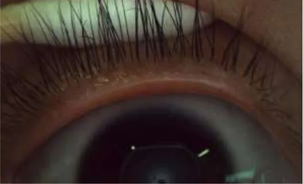
Eyelid Margin
The high-resolution images allow zooming in to closely observe the overall shape of the eyelid margin and detect subtle changes, ensuring thorough and detailed examinations.
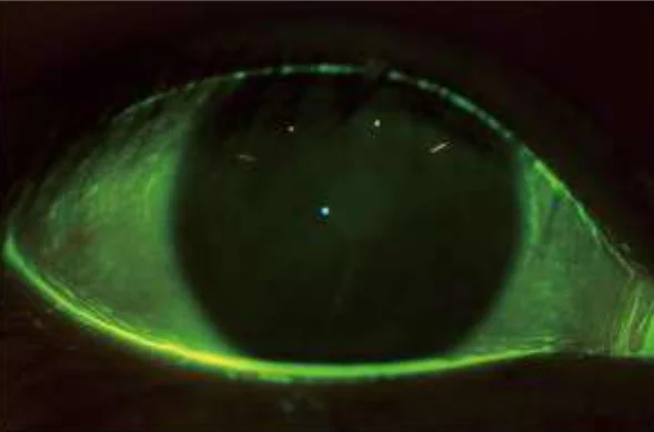
Cornea Sodium Fluorescein Staining
The built-in yellow filter, specially designed to work with cobalt-blue illumination, enhances the contrast of corneal sodium fluorescein images. This effectively improves the detection rate of early corneal epithelial staining, ensuring more accurate and reliable diagnoses.
Corneal Topography
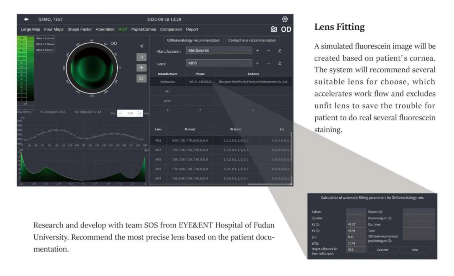
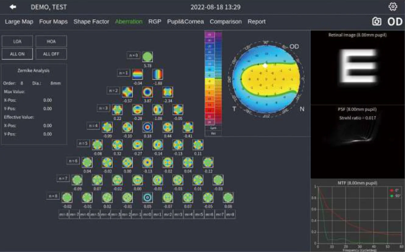

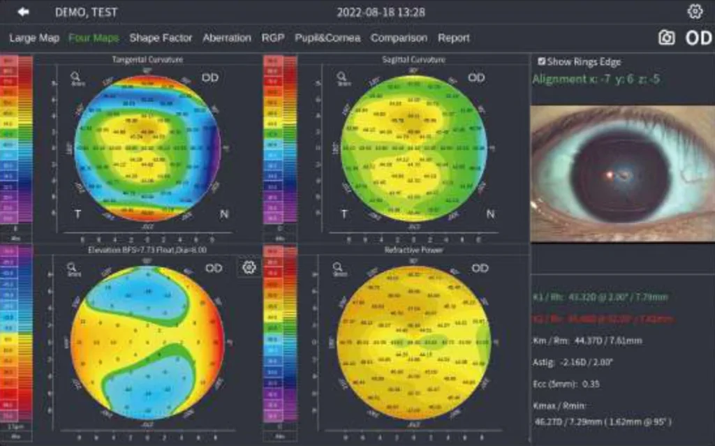
4 Maps
4 maps provide Sagittal Curvature, Tangential Curvature, Elevation Map, Refractive Power, and K1/K2/Km/Astig/Ecc value.
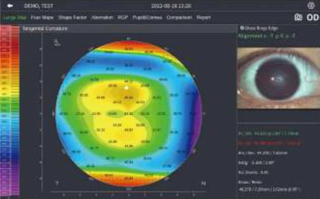
Topography
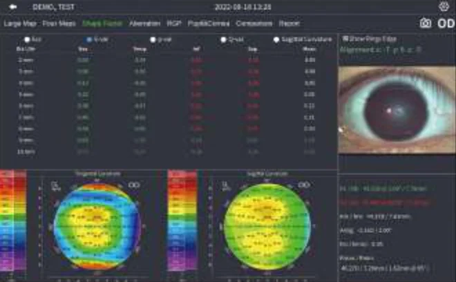
Shape Factor
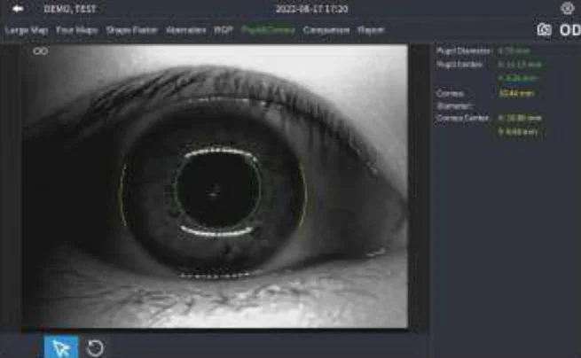
Pupil & Corneal Diameter Measurement
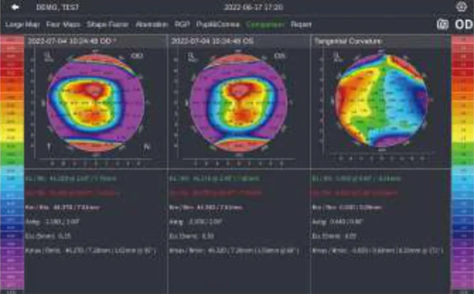
Cases Comparison
Technical Specification ETD1 Ocular Diagnostic Master
| Specifications | Details |
|---|---|
| Hardware | |
| Dimension | 53cm x 30cm x 54cm |
| Weight | 12.79kg |
| Built-in CPU | Intel |
| Hard Disk | 1TB |
| Image Resolution | 2048 x 1536 |
| Display | 10.1" touchscreen |
| Illumination | White, Infrared, Cobalt blue |
| Internet Connection | WIFI |
| Printer Connection | WIFI, USB |
| Power Supply | 100-240VAC, 50/60Hz |
| Extension Display Interface | Display Port |
| OSIOD Recognition | Automatic |
| Chin Rest Control | Electrical |
| Left and Right Work Range | 0-94mm |
| Front and Back Work Range | 0-64mm |
| Up and Down Work Range | 0-30mm |
| Language | English |
| DICOM | Supported |
| Topography | |
| Topography | 50 Rings |
| Diameter of Project Area | 8mm (430) |
| Radius of Curvature | 32.14 dpt - 61.36 dpt (5.5mm - 10.5mm) |
| Accuracy | ±0.1 dpt (±0.02mm) |
| Astigmatism Axis | 0° - 180° |
| White to White | 6mm - 17mm |
| Pupil Diameter | 1mm - 13mm |
| Topography Function | Sagittal Curvature, Tangential Curvature, Elevation Map, Refractive Power |
| 4 Maps | Four Maps Display |
| Shape Factor | P, Q |
| Zernike | Corneal wavefront aberration, PSP map, MTF curve, and Simulated image in different pupil diameters |
| Examination Result Comparison | Support 2 results comparison and difference calculation |
| Dry Eye Analysis | NBUT Automatic analysis, tear film rupture area and time, first break up time, and average break up time |
| Dry Eye Analysis | |
| Nibut | Automatic analysis, tear film rupture area and trend, first break-up time and average break-up time |
| Tear Meniscus Height | 0.01mm - 2mm |
| Meibomian Glands | Meibomian gland loss rate and grade |
| Lipid Layer | Template match |
| Eye Redness | Conjunctival congestion percentage |
| Eyelid Margin | Support digital images zoom in |
| Ocular Surface | Built-in yellow filter |

